Diagnostic care at Michigan Heart Group
At Michigan Heart Group, our team of cardiac specialists is proud to provide a wide range of state-of-the-art diagnostic services with which to evaluate your condition. To learn more about our diagnostic services, explore the links below.
Stress TestingIf you have arthritis or another medical problem that prevents you from exercising during a stress test, you may have the option of taking medicine to make your heart work hard, as it would during exercise. This is called a pharmacological stress test. Stress testing is used to help diagnose heart disease, and to determine how severe heart disease is in people who have it.
Michigan Heart Group has performed more than 50,000 since 1991 and the nuclear lab is accredited by ICANL. The physicians at Michigan Heart Group are highly qualified to interpret your study and make recommendations from it. Specific instructions for each test can be viewed by clicking on the test name below:
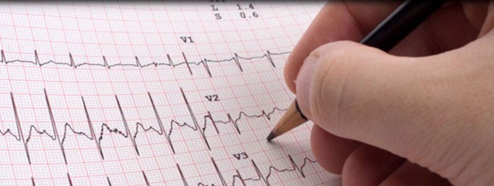
An EKG shows:
- How fast your heart is beating
- Whether the rhythm of your heartbeat is steady or irregular
- The strength and timing of electrical signals as they pass through each part of your heart
Our physicians use EKGs to detect and study many heart problems, such as heart attacks, arrhythmias and heart failure. The test’s results also can suggest other disorders that affect heart function.
A standard surface echocardiogram (TTE) takes images of your heart at rest by placing a probe on your chest. Sometimes images are also taken when contrast is administered through an IV in your arm. A transesophageal echocardiogram (TEE) can be performed by a MHG physician at the hospital if necessary. This test involves swallowing an ultrasound probe under mild sedation. High quality images can be obtained of some structures within the heart that cannot be seen easily from a standard echocardiogram.
Michigan Heart Group has been performing office echocardiograms since 1991. The lab is accredited by ICAEL. The sonographers and physician readers have a vast experience in cardiac echocardiography. Michigan Heart Group physicians have performed over a 1,000 transesophageal studies at Beaumont Hospital since 1991 and were amongst the first physicians in Southeastern Michigan to utilize this procedure.
Specific echocardiogram instructions can be viewed by clicking here.
An event monitor can be worn for weeks at a time. This monitor is activated by the patient or automatically when a symptom or heart rhythm disorder occurs. Home hookups for event monitors can be arranged. Michigan Heart Group has several physicians that specialize in heart rhythm disorders and extensive experience in the interpretation of these tests.
If you have been experiencing symptoms of heart disease or have had an abnormal test result, your MHG cardiologist may want to further evaluate your cardiac anatomy to determine the cause. A cardiac catheterization is one of the most accurate tests to detect the presence of coronary artery disease (blockages) and further indicate which arteries have been narrowed by atherosclerosis. MHG physicians insert a thin, hollow tube called a catheter into an artery in your arm or leg which leads to your heart. The catheter will deliver dye that will show up on an X-ray to visualize the coronary arteries.
If a heart condition is detected, your MHG cardiologist will determine whether medications, angioplasty/stenting, or cardiac surgery should be recommended. The catheterization process also examines leaking or stenosed heart valves, the pumping strength of the heart, and looks for aneurysms and/or heart birth defects. This will help your cardiologist and family doctor determine the best course of action to be taken.
Michigan Heart Group has tremendous experience in performing cardiac catheterization procedures. MHG board-certified Interventional Cardiologists performs hundreds of these procedures for patients with a variety of cardiac conditions.
- Renal Arteries – arteries that supply blood to the kidneys
- Abdominal Aorta – located below the renal arteries
- Iliac Arterie – arteries that supply blood to the leg arteries
- Femoral Arteries – arteries in the legs
- Subclavian Arteries – arteries in the arms
- Carotid Arteries – arteries in the neck
If your physician recommends an:
- Abdominal arteriogram, he will be looking at the blood flow to the renal arteries, abdominal aorta, iliac arteries, and/or the arteries into the legs.
- Arch arteriogram, he will be looking at the blood flow to the great vessels and the vessels into the arm and neck.
Michigan Heart Group Board-Certified Vascular Medicine specialists regularly perform vascular angiography in dedicated vascular labs at Beaumont Hospital with excellent results.
Your MHG physician can tell what kind of heart rhythm problems you have and where those problems are. Sometimes the problem can be fixed at the same time. Michigan Heart Rhythm Group board-certified Electrophysiologists have been performing this procedure for many years and have successfully diagnosed hundreds of patients with heart rhythm disorders.